The brain is a complex object like the universe comprising intricate networks with a tremendous amount of neuronal cells. To understand this structural complexity and its functions, brain atlases that can show fine-scale structures and connections of cellular circuits are a requisite tool for neuroscientists.
Recently, Ueda and his team developed a fluorescent-protein-compatible, intensive tissue-clearing method combined with a tissue expansion protocol, CUBIC-X, which enables seamless imaging of the whole mouse brain. They succeeded in improving the transparency of brain tissue and constructed a point-based mouse brain atlas with single cell annotation (CUBIC-Atlas). Using this 3-D whole-brain atlas, future studies can add activity/gene expression mapping and explore undefined anatomical areas.
The editable CUBIC-Atlas is available to the research community and public (http://cubic-atlas.riken.jp) as a single-cell-resolution platform for unbiased systems-level analysis of mammalian brain.
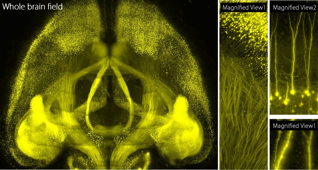
A reconstructed whole brain image based on the acquisition of image data comprising more than 1 million sheets (Whole brain field), Partly-reconstructed brain image data (Magnified View1), Reconstructed image data focusing on neurons (Magnified View2), Reconstructed image data focusing synaptic structure (Magnified View3)
Please watch the movie version below: