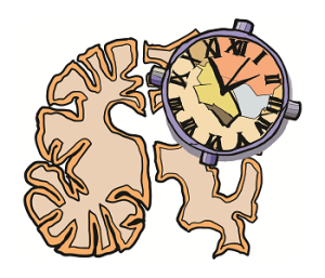
Within the human brain, a region known as the left angular gyrus and the way that it “tracks time” have emerged as a crucial player in the development of Alzheimer's disease.

Tokyo, Japan - Alzheimer’s disease is in many ways like a clock whose time is off, researchers have found. The ‘internal neural timescale’ of a part of the brain is running ‘short’ and throwing off the timing of the rest of a key region.
A paper describing these findings was published in the journal Brain Communications on July 11.
Recent human neuroimaging studies reported atypical anatomical and functional changes in some regions in the default mode network (DMN) in Alzheimer’s patients. The DMN is a collection of brain regions that are active when an individual is at rest or not focused on the outside world. It is often referred to as one of the brain's "resting state networks" because it becomes more active when the mind is wandering, daydreaming, or engaging in internally focused tasks such as self-reflection, envisioning the future, or recalling memories.
The DMN is composed of interconnected brain regions, including the medial prefrontal cortex, posterior cingulate cortex, precuneus, and the angular gyrus. These regions work together to facilitate introspection, social cognition and memory retrieval.
“Disruptions in the DMN have long been implicated in Alzheimer's disease, but the key DMN brain area whose atrophy disturbs the rest of the DMN, and consequently contributes to Alzheimer’s symptoms has until now remained unidentified,” said lead author of the study, Associate Professor Takamitsu Watanabe of the International Research Center for Neurointelligence at the University of Tokyo.
The team examined two types of magnetic resonance imaging (MRI) techniques used to compare the differences in the resting state of the brains of people with and without Alzheimer’s. They made use of functional MRI (fMRI), which captures changes in blood flow and oxygen levels in the brain, in order to allow researchers to observe brain activity and detect which areas of the brain are active during specific tasks. And they also investigated structural MRI data, which provide detailed images of the brain's anatomy, including its size, shape, and identify any abnormalities such as lesions or atrophy. This latter type of imaging is essential for diagnosing neurological conditions and monitoring disease progression.
In this way, the researchers identified a link between the structural changes in the left portion of the angular gyrus, its altered neural activity, and the cognitive decline observed in Alzheimer's disease.
The left angular gyrus is a region located in the parietal lobe of the brain. It is involved in tasks such as reading, writing, language processing, comprehension of complex stimuli, problem-solving and memory retrieval.
Different regions of the brain process time differently, even though some brain areas, such as prefrontal and parietal cortices, integrate these different intrinsic neural timescales (INTs), or “neural clocks,” to integrate diverse information at a single useful timescale. These various neural clocks mediate our behaviors and cognition, and play a decisive role in our sense of self.
Through the comparison of MRI data of Alzheimer’s patients and those without the disease, they found that the neural clock of the DMN in the patients was unusually “short” compared to the cognitively typical subjects.
“When neuroscientists say that a neural clock is running ‘shorter’ or ‘longer,’ we are referring to how quickly or stably neurons in a region hold information within them,” said Shota Murai, another member of the research team.
This atypically short DMN neural clock was also associated with an unusually short neural clock in the left angular gyrus.
This in turn was associated with an unusually low volume of grey matter—the tissue in the brain involved in the integration of diverse information including memory, emotions and movement —in this region.
The researchers believe that the low grey matter volume of the angular gyrus is what shortens its neural clock, which then destabilizes the neural clock of the entire DMN—which contributes to the cognitive decline observed in Alzheimer’s patients.
They hope that tracking such disturbance of the neural clock could be used as a biomarker to detect early onset of Alzheimer’s disease. Better still, they aim to investigate whether stabilizing this neural clock could slow down progression of the disease.
###
The article, “Atypical intrinsic neural timescale in the left angular gyrus in Alzheimer's disease”, was published in Brain Communications at DOI: 10.1093/braincomms/fcae199.
JOURNAL
Brain Communications
DOI
10.1093/braincomms/fcae199.
METHOD OF RESEARCH
Imaging analysis
SUBJECT OF RESEARCH
People
ARTICLE TITLE
Atypical intrinsic neural timescale in the left angular gyrus in Alzheimer's disease
ARTICLE PUBLICATION DATE
11-Jul-2024


