A new study in mice has revealed never-before-seen details about how the complicated visual network forms in them. This research could inform future research into the treatment of congenital blindness. But given the parallels between biological neural tissue and digital artificial intelligence, this research could also help software engineers develop better and more general-purpose artificial intelligences.
If you could see the weblike nature of the neurons and structures that make up the brain and sensory systems of animals, you might think it’s just a random complicated mess. But researchers such as neuroscientists are able to look at this chaos and deduce not only discrete structures, but also ascertain their functions. Recently, Professor Kenichi Ohki, Assistant Professor Tomonari Murakami, and colleagues from the International Research Center for Neurointelligence (IRCN), Institute for AI and Beyond and the University of Tokyo School of Medicine have been studying a particular brain network to learn how it forms — the visual system.
“The eyes, certain parts of the brain and the neural network connecting these form the vision system. A crude analogy might be a camera connected by a wire to a screen that your conscious self can watch. But an accurate biological description of this system is extremely complicated,” said Murakami. “There is a large number of visual cortical areas involved and these are arranged in layers which form a sort of hierarchical structure. This idea is not new, but it was not known how connections between the early stages of this network, or primary areas, and areas involved in the processing of visual signals, or higher visual cortical areas, form during development. We set out to find out how this happens.”
The team studied the developing visual system of mice. In particular they looked at areas called cortical and thalamic regions. By seeing how networks of neurons in these regions developed in newborn mice, and when these networks became active, the team was able to describe in a more general way the mechanisms governing the growth of the visual system.
“As we recorded the increasingly dense network of connections in time, something jumped out that surprised us,” said Murakami. “We expected the visual network to form a lot of connections among the cortical areas first, reflecting the hierarchical structure of the whole system. But, in fact, parallel neural pathways from the retinas in the eyes leading up to the cortical areas form earlier than those among cortical areas. This new fact changes what we know about this area of cortical development.”
This study was done not only to satisfy curiosity, but also because fundamental research of this kind can form the foundation of future medical research which can improve peoples’ lives: in this case, the team’s hypothesis that their research in mice can likely explain visual development in primates, including humans. And this in turn could help researchers aiming to treat congenital blindness.
“There is another field of research that can learn from what we have done here as well,” said Ohki. “Artificial intelligence is often based on digital artificial neural networks. These are usually structured in multiple layers, which can give them complex functionality. But now that we’ve shown at least some biological neuronal systems develop parallel structures prior to layered ones, software engineers might gain inspiration from this to experiment with new design methodologies. It is conceivable this might help them in their goal of creating ever more general-purpose intelligences capable of solving a wide variety of problems.”
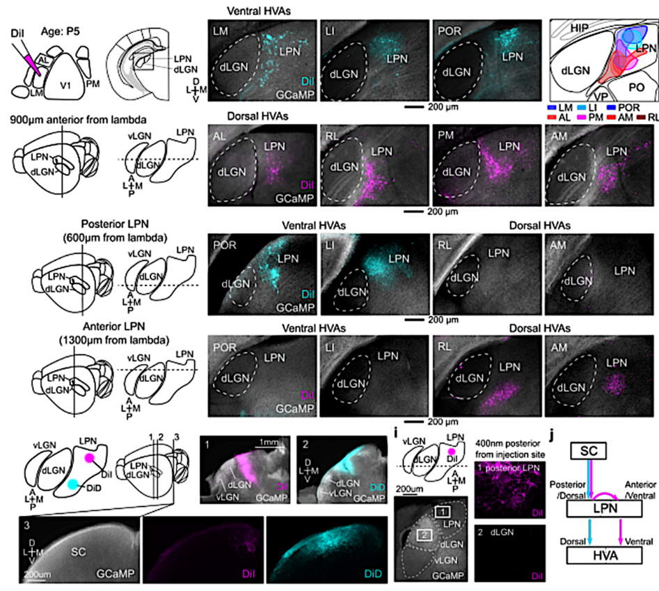
Visual networks in developing mice. These scans of a developing mouse brain show that higher-order cortical visual areas receive visual information from thalamic nuclei. The magenta and cyan colors represent different neuronal pathways that form earlier than connections within the cortical areas. ©2022 Murakami et al.
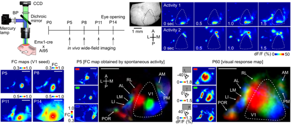
Developmental change. The researchers were able to visualize developmental changes in the mouse visual system. The patterns shown in visualizations such as these show the spatiotemporal pattern of neural activity in the cortical visual areas. ©2022 Murakami et al.
Journal article: Tomonari Murakami, Teppei Matsui, Masato Uemura, Kenichi Ohki. “Modular strategy for the development of hierarchical networks in the mouse visual system”, Nature. DOI: 10.1038/s41586-022-05045-w
www.nature.com/articles/s41586-022-05045-w
Funding: This work was supported by Brain/MINDS-AMED (JP16dm0207034, JP20dm0207048 to K.O.); Institute for AI and Beyond (to K.O.); JSPS KAKENHI (25221001, 19H05642, 20H05917, to K.O., 17K14931, 18H05116, 21H0516513 to T. Matsui, 19K21207, 21K15181 to T. Murakami); Brain/MINDS-beyond-AMED (JP20dm0307031 to T. Matsui); and JST PRESTO (JPMJPR19M9 to T. Matsui).
Useful links:
World Premier Institutes - International Research Center for Neurointelligence (WPI-IRCN)
Graduate School of Medicine
Institute for AI and Beyond
Research contact: Professor Kenichi Ohki
Press contact:
Mr. Rohan Mehra